The latest paper for #microtwjc focuses on finding a new way to sense metabolites inside of living bacteria. Bacteria need to regulate their nutrient intake, because like people, they don't want to get too fat or thin.
If there isn't enough nutrient in the environment, they need to conserve what they have, and if more appears, they need to consume it as efficiently as possible, but if they did that all the time, it could cause them problems.
But we don't have many great ways of observing how bacteria do this. There is a need for the creation of biosensors, which can sit in the bacteria without killing them and tell us what they are doing with their nutrients in whatever situation they find themselves. In this paper, they were looking at the localisation of a particular nutrient, citrate. So how do you do this? where do you find these sensors? Luckily, mother nature's got you covered.
Lots of bacteria have proteins which are used to grab citrate from the environment, like a periplasmic binding proteins which pretty much do exactly that. Bacteria also have "sensor kinases", which essentially work as sensors for the bacteria themselves, so that they know how much nutrient they themselves have. All of these proteins bind citrate. When they do bind, they change their shape.
But we can't just put them under a microscope, because these proteins are too small to see, and the only microscopes powerful enough to work would kill the bacteria. So what's a microbiologist to do ?
Don't fret ! FRET will come to the rescue ! What is FRET, and why am I shouting about it ?
FRET is an acronym, which stands for Fluorescence Resonance Energy Transfer. There, I've explained it, no more need to dwell on... okay fine. Let's quickly go through what Fluorescence Resonance Energy Transfer is.
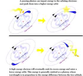
Essentially, fluorescence is when you use photons to excite electrons that are bonded to atoms. Every electron has a specific orbit around an atom. This orbit relates to the level of energy it possesses.
If you shoot a photon of a specific wavelength, it will cause the electron to go into a higher energy state for a while. and then the electron will jump back down , releasing a photon.
Not just any photon will do either, it has to have a specific frequency. A molecule that fluoresces (which we're calling a fluorophore) under UV light will absorb ultraviolet radiation in order to get its electrons excited. But the light it emits afterwards is usually of a lower frequency, in this case it's blue light.
There are a lot of biological fluorophores around, most of them derived from green fluorescent protein. These can be encoded onto DNA and inserted into cells to make them glow when excited by specific light.
So that's Fluorescence sorted out. What about Resonance Energy Transfer?
Here, we'll need two fluorescent proteins. One of them absorbs say absorbs violet light, and emits blue light, and the second absorbs blue light and emits yellow light.
So if we shine just the violet light, we'd see blue light from the first protein, and none from the second one. But if we bring those two fluorophores close together, this changes.
In simple terms, they "get in eachothers bizniss".
In the excited fluorophore, we have electrons in a high energy state. But they interact with the electrons in the other fluorophore, and end up transferring their energy to them in order to get into a lower ground state. This is done via a "virtual photon" that barely has time to exist before exciting the other fluorophore.
So this way, you can shine violet light in a mixture of these two fluorphores. And you'll be able to tell how close they are to eachother by the light you get back off of them. If you get yellow light, they are close, and if you get blue light they are not. In fact the proportion of yellow light compared to blue light produced relates to how distant these fluorophores are from eachother.
But it also gets more subtle. So let's get back to the citrate binding proteins, and how we detect them changing shape when they bind to citrate.
If you attach a fluorophore to one end of the protein and another to the other end, then if the shape of the binding protein changes, so should the distance between the fluorophores. Essentially, you have created a biosensor. It will emit a different spectrum of light depending on whether it has bound to citrate or not.
So now we've gone through some of the basic concepts of the paper, let's start going through the paper.
Figure 1
The first thing they did was to try out a number of different citrate binding proteins. For their fluorophores, they decided to use Cyan Fluorescent Protein, and Venus, which is a derivative of Yellow Fluorescent protein.
They tried out a number of different citrate binding proteins. They had no idea which ones would work, or even why they'd work. The basically worked things out empirically, which is short hand here for "try out everything until something works"
In they end, they found that the CitA protein from Klebsiella was the one which worked. And by worked, I mean that it showed some difference in wavelength when citrate was added.
The bottom axis is the wavelength of light, with 480 being violet/blue and 540 being around the red/yellow part of the spectrum. The fluorescene intesnity is basically how bright the light is coming off of the sensors.
The dotted line is the protein without the citrate added, and the black one is when it has been added.
When citrate is added, more light is emitted from the blue spectrum, and less is emitted from the yellow spectrum, implying (if i've got my physics right) that the fluophores have slightly moved further from eachother.
Using this, you can calculate the fluorescence intensity ratio, by looking at the amount of yellow (530) light produced divided by the amount of blue (488) light produced. If there is no change, then the ratio stays the same. If the fluorophores move apart, then this the light at 488 decreases, and 530 increases, and fluorescence intensity ratio get's much higher.
But this change is still small, which would could produce detection problems.
Figure 2
The next thing the researchers did was to try and make these small changes in wavelength bigger. They looked at the protein structure of CitA, and removed flexible bits, as the "wobbling" of a protein will make the distance between the fluorophores vary, and make it more difficult to tell whether any shape change is due to binding of citrate, or due to the proteins natural wobbliness.
In they end, they eliminated two sections of the protein, and found that this improved their signal.
So what next ?
Next, they want to look at how sensitive their biosensor was at detecting citrate.
A : They added different concentrations of citrate, and then marked up the fluorescence intensity ratio. They then looked at how much the signal increased in response to adding more citrate. What you can see is a basic enzyme binding curve. Essentially, it shows that this biosensor cannot really detect concentrations of citrate lower that 1 micro mol, and that it is less sensitive at detecting concentrations in excess of 100 micromols.
B: But it's not enough to have a sensor that is very sensitive, it has to be specific. So they added a variety of different metabolites to this sensor,t o see if they made any difference. Only one did, an isomer of citrate, isocitrate. In this case, faint detection only occurred at around 10-1000 micro mols, far more than you ever expect to see in a cell. They also mixed together isocitrate with the citrate to check if there was any binding. while they don't show the actual data, they say that no competition is happening.
Table 1
I noted before that the biosensor was less sensitive when the amounts of citrate exceeded 100 micro molar. This is not great, as in a real cell, the sensor might have to deal with concentrations that high. So they did some more engineering on the CitA protein, to alter the way it binds so that it could detect higher concentrations of citrate.
The way they had to do this was to make the CitA bind less well to citrate. This would mean that the concentration of citrate would need to be higher in order for binding to occur.
They introduced a few mutations into CitA to do this, and then repeated the above experiment. They only display the KD as this basically shows the halfway point, where the area where the most sensitive detection can occur lies on the above graph.
The below table shows the binding properties of six mutated versions of the biosensor (CIT8u is the name of the normal biosensor)
They combined the mutations for CIT50u and CIT96u to create sensor with an affinity for 470 uM of citrate.
So now they had a panel of different biosensors which could cover a wide range of concentrations found in the living cell. So how can we tell which ones work ?
Figure 3
The best way for them to test these sensors was to try the out in living cells. They put the genes for these biosensors into a lab strain of E. coli. They used the CIT8u, CIT96u, CIT1.8m, to check which one would be the most appropriate. They also included a sensor which didn't bind anything, CIT0, as a control.
They were grown at room temperature for 60 hours, and then starved of nutrients on M9 media for four hours. After this, they then either added, glucose, acetate or citrate to the cells at a concentration of 2ug/ul.
So the first thing to address here is which sensor works ? Well, none of them really. When using biosensors properly, it's quite difficult to gauge how sensitive they are because you are relying on the bacteria to make them themselves. They don't really know how much biosensor a bacterium is going to make, and whether they all make the same numbers of biosensors per cell. They don't mention the plasmid they used for this study, so I cannot tell what kind of copy number to expect from it, or how active the promoter is for the biosensor.
While it will likely hover around the same average, this variation can throw out the kind of sensitive analysis performed in the in vitro experiments. So the only clue to the amount of citrate present comes from using sensors of different affinities, and seeing how well they compare to eachother.
Now we can get into what the data shows- firstly, when bacteria metabolise glucose, citrate is produced as the end point of glycolysis. Citrate build up for a time, and then falls as the TCA cycle starts up and begins to metabolise the excess citrate.
When Acetate is metabolised, citrate levels are increased for a bit longer, as citrate is one
Whereas when citrate is added, the E.coli do not absorb it, as it tends only to be absorbed by these bacteria under anaerobic conditions.
So now that they've seen how these biosensors can detect citrate when it is produced during the metabolism of glucose and acetate, they wanted to see whether they could find out anything new with their fancy new biosensors.
Figure 4
They took E.coli cells containing the CIT8u biosensor, and then starved them for 24 hours. At the end of these 24 hours, they added either glucose or acetate, and measured how quickly they were metabolised to citrate in the E.coli cells.
Turns out that both of these get metabolised very quickly, with the change due to glucose uptake (in black dots) being very rapid and similar results for acetate. This suggests that glucose uptake is very quick after 24 hours. But how do we know it's quicker than uptake after different levels of starvation?
In this graph, we have measurements taken every ten seconds being compared to measurements taken every minute. Will this make much difference to the results ? Probably not.
But it would have been nice to see how uptake of glucose/acetate changes at different degrees of starvation using directly comparable data sets. You could use statistics to compare the data, and perhaps reveal something interesting about how quickly bacteria adapt to starvation.
Summary
The Good
I couldn't find much wrong with this paper when it was dealing with the basic engineering issues for creating the citrate biosensor.
The focus was on getting a product that works. The work needed to find the crystal structures for all of these biosensors is huge, and not necessary. We don't need to know the exact mechanics, all we want is something that can bind citrate, and then give us the signal that it has.
They have pretty much shown that they have made a series of biosensors that are specific to citrate, and sensitive over a wide panel of concentrations. The methods they use when creating their biosensor are really good, and made an interesting read.
The Bad
Where the paper slightly falls down is when they try to prove that their sensors work in real living bacteria. Did they do enough to work out whether their biosensor is being expressed well enough ?
Did the biosensor inflict any fitness cost on the bacteria carrying it ? How stable is it on minimal and rich media? If I grow a batch of E.coli with it, will they still express it in a week ?
These are vitally important questions for the application of biosensors like this one. If you take one reading at say 4 hours, and another at 24 hours and half the cells may have already spat out your plasmid, and you'll be none the wiser if you only look at the fluorescence ratio, other than noticing that your data gets more variable the longer you leave your bacteria.
The data presentation left me annoyed. Some of the data was presented really well, other times not so much. Sometimes you get to hear how many replicates happened in each experiment, sometimes you won't. You may see error bars in one, and they are gone in the next experiment.
Sometimes they appear, and are not explained. If you don't mention what the error bars mean on a graph, then they can only be judged as pretty little lines.
Verdict
The thing about this paper is that it's got a lot of good stuff going for it, and only starts getting shaky when we leap into working in living bacteria. They haven't fully characterised their sensor in living bacteria. But even if I let that slide, we still have problems with data presentation that make it difficult to work out what they actually did. It's a small point, but an important one if a scientist like me needs to implement a biosensor like the one described here.
Despite this, there is still enough good stuff here for me to have some respect for this work. I am pretty much convinced that they've created an interesting set of biosensors that could potentially be useful. It's a great paper, but not perfect.
(2011). Engineering Genetically Encoded Nanosensors for Real-Time In Vivo Measurements of Citrate Concentrations, PLoS One, 6 (12) DOI: 10.1371/journal.pone.0028245.t002


Fantastic explanation! You had me at fatty bacteria :P
ReplyDelete